
Cranial Nerves and Spinal Nerves: Why we have both.
Why Do We Have Cranial Nerves
AND Spinal Nerves?
I was recently asked a brilliant question: why do we need cranial nerves as well as spinal nerves and why can’t one take over if the other one gets damaged? Let’s give it a go!
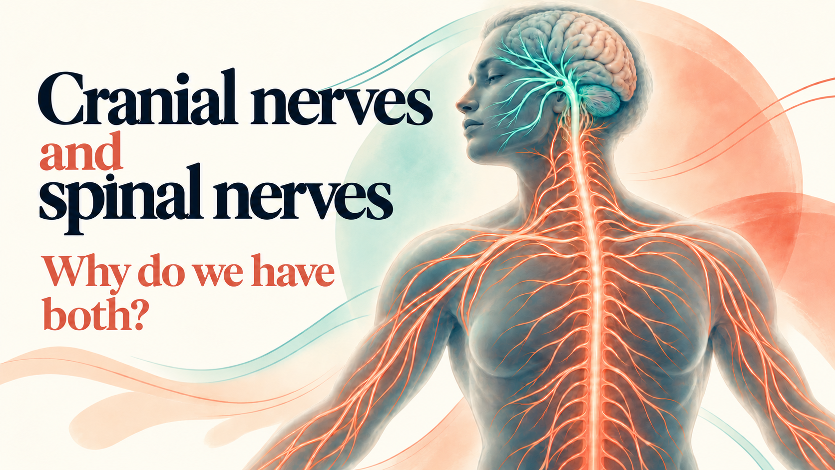
We have 31 pairs of spinal nerves connecting the spinal cord with most of the body and 12 pairs of cranial nerves associated with the brain and brainstem. Some (in both sets) are sensory, some are motor and some are both. But if both can carry sensory and motor information, why have two arrangements at all? And if a cranial nerve can make a muscle contract (which it can), why couldn't it take over when the spinal cord is damaged?
This is all about how the nervous system is organised as cranial and spinal nerves aren't two separate nervous systems. Instead they are different routes connecting different parts of the central nervous system with particular areas and functions, something that becomes significant when something goes wrong.
Most of the trunk and limbs communicate through the repeated, segmental arrangement of the spinal cord and spinal nerves, (lumbar thoracic, cervical etc.) (Kaiser and Lugo-Pico, 2023). The head, meanwhile, contains specialised sensory organs and muscles concerned with things including vision, hearing, balance, facial sensation, swallowing and facial expression, with many of those pathways connecting directly the brain and brainstem. (Sonne, Omole and Lopez-Ojeda, 2025)
The distinction isn't completely tidy, because biology rarely is. The vagus nerve leaves the skull and travels into the chest and abdomen, while the accessory nerve, traditionally classified as a cranial nerve, has a significant relationship with the upper spinal cord and supplies two muscles very familiar to therapists: sternocleidomastoid and trapezius.
Most of us at some point with with heads, necks, jaws, shoulders, breathing, balance or movement, so cranial nerves are worth knowing a bit about as they provide a useful reminder that where somebody experiences something, isn’t necessarily where the story begins.
Spinal nerves.Relatively well behaved
Our 31 pairs of spinal nerves connected to the spinal cord contain sensory and motor fibres and act as pathways between the central nervous system and the rest of the body (Kaiser and Lugo-Pico, 2023).
Sensory information travelling towards the central nervous system is generally described as afferent, while motor information travelling away from it is efferent. My way of remembering the difference is that effluence — sewage — needs to be taken away. Whatever works right?
Then comes the twist! As nerve fibres travel towards the limbs via the spine, fibres from different spinal levels mix together in plexuses before being redistributed into peripheral nerves. The median nerve in the arm or the sciatic nerve in the leg aren’t simply a cables plugged into one particular bit of spinal cord, but contain fibres with different spinal origins, all travelling together (Kaiser and Lugo-Pico, 2023).
The head’s wiring arrangements
Function is key to learning cranial nerves, rather than memorising cranial nerve names (although I have included a list) and looking for common functions is the starting point.
Clench the jaw and the trigeminal nerve, cranial nerve V, is involved. It supplies the main muscles of mastication and carries sensory information from the face. Smile or raise the eyebrows and the facial nerve, VII, supplies the muscles producing those expressions. Moving the eyes involves three different cranial nerves, III, IV and VI, while the vestibulocochlear nerve, VIII, brings information from the inner ear concerned with hearing and balance. Stick the tongue out and XII, the hypoglossal nerve, shows up (Sonne, Omole and Lopez-Ojeda, 2025).
And then there’s the shoulder! The accessory nerve is my favourite example because it messes nicely with the categories. Ask somebody to shrug their shoulders or turn their head against resistance and two familiar muscles become involved: trapezius and sternocleidomastoid. Many therapists probably don’t realise that these muscles’ primary motor supply comes from a cranial nerve accessory nerve XI (Sonne, Omole and Lopez-Ojeda, 2025).
The story gets more interesting because the spinal part of the accessory nerve arises from the upper cervical spinal cord before entering the skull and then leaving it again! (Sonne, Omole and Lopez-Ojeda, 2025). Tidy anatomy tends not to respect the boundaries of book chapters!
Balance isn’t just strong ankles
Cranial nerves also give us a useful way of thinking about movement
Standing on one leg can be seen as a muscular task involving ankle stability, gluteal strength, foot position or trunk control, but staying upright also requires a considerable amount of sensory information.
The brain continually combines information from vision, the vestibular system of the inner ear and sensory information from muscles, joints and skin. Together these systems help us establish where the body is, how the head is moving and what needs to happen to maintain posture and balance (National Institute on Deafness and Other Communication Disorders, 2018).
If balancing with the head turned or the eyes closed changes things, nothing has changed in the muscular system.Instead the information available to the nervous system has changed, along with what it’s being asked to do.
Information detected in the vestibular apparatus about head position and movement is carried centrally to the vestibular nerve VIII.This is then integrated with information from the visual cortex as well as somatosensory information from the spine, all contributing to the idea of balance, orientation and vision during movement. (National Institute on Deafness and Other Communication Disorders, 2018).
This is a huge deal for therapists, particularly movement practitioners. Someone who appears perfectly stable during one version of a movement but becomes less so when head position changes, may be showing us something more than a weak muscle. We’re not able to diagnose a cranial nerve disorder, but we are able to ask better questions and avoid chasing dead ends.
The face can tell us quite a lot
The same principle applies when somebody arrives with an apparently ordinary neck problem. Changes in facial sensation, chewing, hearing, swallowing or speech, should change how we think about the presentation as all these functions involve several different cranial nerves.(Sonne, Omole and Lopez-Ojeda, 2025).
If someone has neck discomfort but also changed facial sensation, for example, avoid assuming that both things come from the neck.
Similarly dizziness associated with head movement might have several possible explanations and deserves more consideration than thinking of a cervical spine approach. My biggest lessons over the years have come from listening to the feeling that the presentation doesn’t quite fit the musculoskeletal story and digging more.
And inevitably, the vagus nerve
It’s almost impossible to talk about cranial nerves nowadays without mentioning the superstar celebrity of the group, winner quite literally of the X factor, the vagus nerve, cranial nerve X.
It’s an unusual and genuinely fascinating bit of anatomy because it doesn’t just stay conveniently around the head and neck, but travels into the thorax, abdomen and beyond. It also has a wide range of roles to play, including sensory, motor and autonomic functions involving structures including the pharynx, larynx, heart, lungs and digestive tract. (Sonne, Omole and Lopez-Ojeda, 2025).
This range of tasks also makes it extraordinarily marketable, with vagus nerve resets, vagal release techniques and various ways of supposedly manipulating it being widespread in therapy and wellness circles.
It’s a this point that we need to separate ‘interesting anatomy’ from ‘demonstrated outcome’. Someone may feel calmer after touch, movement, breathing or spending an hour with another human being who cares.That experience is incredibly valuable. BUT, this feeling doesn’t automatically demonstrate that somebody’s fingers have ‘reset the vagus nerve’.
This distinction between observation and explanation is something that often gets blurred in our industry. ‘I feel calmer’ describes an experience. ‘I manually stimulated your vagus nerve and switched your nervous system into parasympathetic mode’ proposes a biological mechanism. The first doesn’t prove the second, however attractive it might sound.
Knowing when the map has changed
Sometimes changes in function have a more urgent meaning. Sudden facial weakness, arm weakness or speech disturbance are classic signs of stroke and require emergency action even if it seems the event has passed. The NHS FAST message remains a useful and deliberately simple way of recognising some of the common signs: Face, Arms, Speech, Time to call 999 (NHS England, 2024).
We don’t need to identify cranial nerve VII or locate a lesion somewhere in the brainstem before deciding that something is wrong. Simply recognising that there has been a significant neurological change is enough.
Spinal and cranial nerves give us different ways of mapping the same nervous system, but at the end of the day, neither map is the person sitting in front of us and knowing the names structures doesn’t help in finding out the real story of who that person is.
References
Kaiser, J.T. and Lugo-Pico, J.G. (2023) ‘Neuroanatomy, spinal nerves’, StatPearls. Treasure Island, FL: StatPearls Publishing. Available at: https://www.ncbi.nlm.nih.gov/books/NBK542218/ (Accessed: 5 October 2026).
National Institute on Deafness and Other Communication Disorders (2018) Balance disorders. Bethesda, MD: National Institutes of Health. Available at: https://www.nidcd.nih.gov/health/balance-disorders (Accessed: 5 October 2026).
NHS England (2024) NHS launches major new stroke campaign as thousands delay calling 999 by nearly 90 minutes. 4 November. Available at: https://www.england.nhs.uk/2024/11/nhs-launches-major-new-stroke-campaign-as-thousands-delay-calling-999-by-nearly-90-minutes/ (Accessed: 5 October 2026).
Sonne, J., Omole, A.E. and Lopez-Ojeda, W. (2025) ‘Neuroanatomy, cranial nerve’, StatPearls. Treasure Island, FL: StatPearls Publishing. Updated 24 January 2025. Available at: https://www.ncbi.nlm.nih.gov/books/NBK470353/ (Accessed: 5 October 2026).


